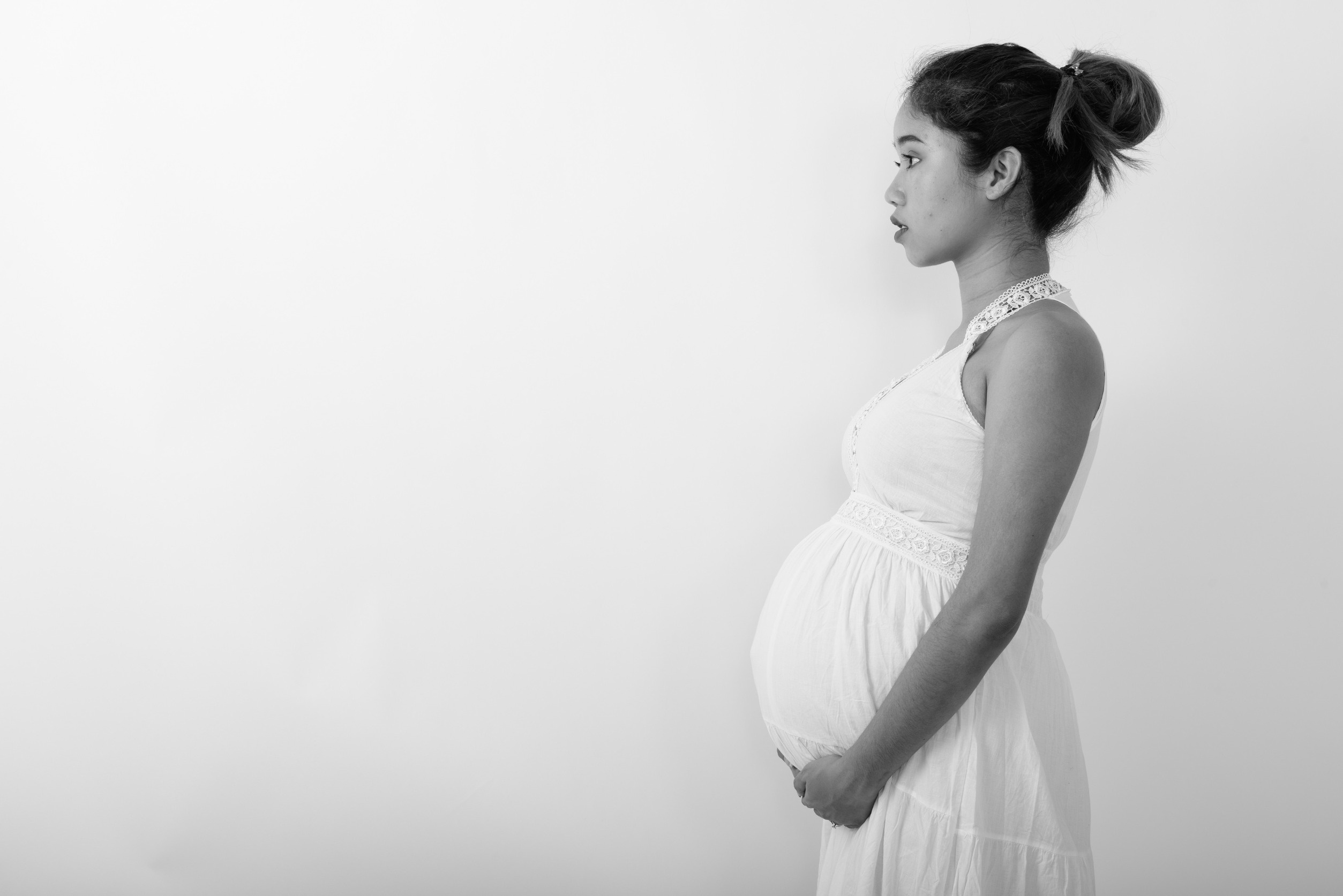
Did you know pregnancy changes the brain? Researchers have created one of the first comprehensive maps of how the brain changes throughout pregnancy, substantially improving upon understanding of an understudied field.
Certain brain regions may shrink in size during pregnancy yet improve in connectivity, “with only a few regions of the brain remaining untouched by the transition to motherhood,” according to the study published Monday in the journal Nature Neuroscience.
The findings are based on one healthy 38-year-old woman the authors studied from three weeks before conception to two years after her child’s birth. Dr. Elizabeth R. Chrastil, a professor at the University of California, Irvine, underwent in vitro fertilization. Chrastil conceived the project and wished to use herself as the participant, as has been done in previous research.
There has been “so much about the neurobiology of pregnancy that we don’t understand yet,” said senior study author Dr. Emily Jacobs, associate professor in the department of psychological and brain sciences at the University of California, Santa Barbara, in a news briefing on the study. “And it’s not because women are too complicated. … It’s a byproduct of the fact that the biomedical sciences have historically ignored women’s health. It’s 2024, and this is the first glimpse we have at this fascinating neurobiological transition.”
“The brain is an endocrine organ, and sex hormones are potent neuromodulators, but a lot of that knowledge comes from animal studies,” Jacobs said. Human studies tend to rely on brain imaging and endocrine assessments collected from groups of people at a single point in time.
“But that kind of group averaging approach can’t tell us anything about how the brain is changing day to day or week to week as hormones ebb and flow,” Jacobs added. “My lab here at UC Santa Barbara uses precision imaging methods to understand how the brain responds to major neuroendocrine transitions like the circadian cycle, the menstrual cycle, menopause and now, in this paper, one of the largest neuroendocrine transitions that a human can experience — which is pregnancy.”
How pregnancy changes the brain
Jacobs and colleagues conducted 26 MRI scans and blood tests on the first-time mother, then compared them with brain changes observed in eight control participants who weren’t pregnant.
• By the ninth week of pregnancy, the authors found widespread decreases in gray matter volume and thickness of the cerebral cortex, especially in regions such as the default mode network, which is associated with social cognitive functions.
• Gray matter is an essential brain tissue that controls sensations and functions such as speech, thinking and memory. After peaking during childhood, cortical thickness decreases throughout one’s lifespan.
• The scans also showed increases in cerebrospinal fluid and white matter microstructure in the second and third trimesters, all of which were linked with rising levels of the hormones estradiol and progesterone..
• Some of the changes — including cortical volume and thickness — remained two years after birth, whereas others reverted to levels similar to those of the preconception period by roughly two months postpartum.
• And compared with the control group, change in the woman’s gray matter volume was nearly three times higher.
“This study is fundamental in laying the groundwork for future research by providing data that allows future research to explore in more detail and look at in relation to how we can support healthy brain changes in pregnancy (in the mother which will likely impact that developing fetus),” said Dr. Jodi Pawluski, a neuroscientist, therapist and author based in France, via email. Pawluski wasn’t involved in the research.
What brain changes mean for parents
The functional implications these brain changes may have for birthing parents have yet to be determined, said Dr. Elseline Hoekzema, head of the Pregnancy and the Brain Lab at Amsterdam University Medical Center, via email. Hoekzema wasn’t involved in the study.
• However, some of Hoekzema’s previous work has indicated associations between pregnancy-related brain changes and the ways a birthing parent’s brain and body respond and bond to infants’ cues, Hoekzema added.
• These findings are also in line with animal studies showing brain changes that were critical for the onset and continuation of maternal care.
• The decreases in gray matter volume and cortical thickness could point toward an idea that for the maternal brain, “it looks like less really may be more,” Pawluski said. “It’s potentially becoming more efficient.”
• The increase in white matter microstructure, on the other hand, could mean “an increase in the exchange of information and communication between different brain areas,” Pawluski said. The findings may also have important implications for preventing or treating perinatal mental health issues or supporting a healthy transition to motherhood.
• Research thus far shows the changes “are relatively very consistent across different women,” Hoekzema said. “In one study we found that all the participants could be classified as having been pregnant or not by a computer algorithm based only on the changes in their brains. And so far we’ve already replicated these changes.”
• Despite the unanswered questions, Pawluski said she wants birthing parents to know these changes are normal and healthy, rather than a deficit that some people believe is a stereotypical experience of motherhood.
Click here to read more on how pregnancy changes the brain.


